טיפות דמעות – מה זה ולמה משתמשים בהן?
טיפות דמעות הן תמיסות רפואיות או קוסמטיות שמטרתן להקל על


Pterygium, a common disorder of the ocular surface, is characterized by a fibrovascular tumor invading the cornea from the conjunctiva. This article provides an in-depth scientific review of the pathogenesis, epidemiology, clinical manifestations, diagnosis and treatment of pterygium. It also offers insights into current research directions and future treatments in this area.
Pterygium is a chronic and progressive disorder of the eye surface that is especially common in populations exposed to high levels of ultraviolet (UV) radiation. It is often considered a benign condition, however, its progression can lead to visual impairment, cosmetic problems and recurrent infections.
Pterygium mainly affects adults over the age of 40, with a higher incidence in areas closer to the equator. Pterygium is associated with increased exposure to UV radiation, work in an outdoor environment, male gender and older age. Its prevalence ranges from 0.7% to 33% worldwide, depending on the geographic location, climatic conditions and the population being examined.
Although exposure to UV radiation is believed to be the primary cause, the exact pathogenesis is still not fully understood. There is a strong relationship between UV-induced oxidative stress and inflammation in developing pterygium. Oxidative stress leads to damage and mutations in DNA, which contributes to cell proliferation, inflammation and angiogenesis, an integral part of pterygium development.
Emerging evidence suggests that limbal stem cell deficiency, viral infections, such as human papillomavirus (HPV), and genetic predispositions may also contribute to the pathogenesis of pterygium.
Patients with pterygium may be asymptomatic in early stages. However, as the condition progresses, symptoms can include irritation, redness, tearing, foreign body sensation, and blurred vision. The most common sign is a fleshy, triangular, fibrovascular tumor extending from the conjunctiva to the cornea, usually on the side of the nose.
The diagnosis is mainly clinical, based on the characteristic appearance of the lesion. For atypical lesions or suspected malignancy, such as ocular surface squamous neoplasia (OSSN), histopathologic examination may be warranted.
The main treatment for pterygium is surgical removal. However, due to high recurrence rates (up to 88% in some studies), multiple techniques have been developed to reduce recurrence, such as conjunctival membrane transplantation, amniotic membrane transplantation, and the use of adjunctive therapies such as mitomycin C and bevacizumab.
Recent advances include minimally invasive pterygium surgery and the use of fibrin glue instead of sutures. However, these techniques need further validation in terms of long-term safety and efficacy.
Recent studies have focused on the molecular biology of pterygium to develop targeted therapies. This includes studying the roles of growth factors, cytokines, matrix metalloproteinases and vascular endothelial growth factor (VEGF).
Future studies are expected to explore gene therapy, stem cell therapy, and new drug treatments. In addition, research on UV protection strategies, such as promoting the use of UV-blocking sunglasses, can contribute to prevention efforts.
Pterygium, although often considered a benign condition, can lead to significant discomfort and visual impairment. A better understanding of its pathogenesis, combined with advances in surgical techniques and new treatments, is essential to improving patient outcomes. Future research directions are promising, with significant potential for the development of more effective and targeted treatments and prevention strategies.
Author: Prof. Michael Mimouni
טיפות דמעות הן תמיסות רפואיות או קוסמטיות שמטרתן להקל על
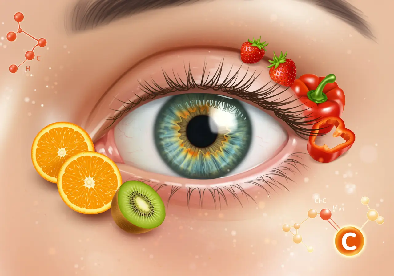
ויטמין C הוא אחד הוויטמינים החיוניים ביותר לבריאות העיניים, הודות