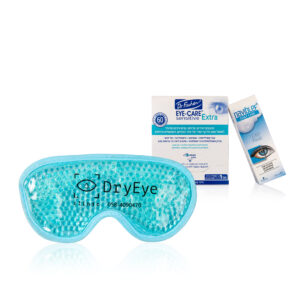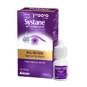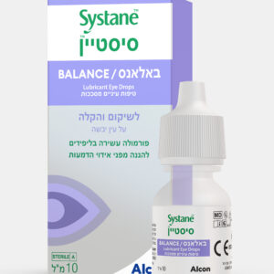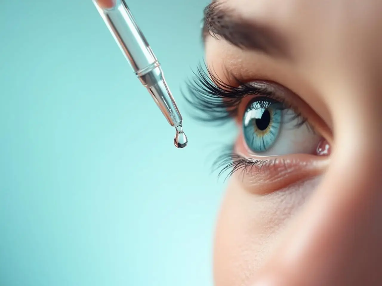
טיפות דמעות – מה זה ולמה משתמשים בהן?
טיפות דמעות הן תמיסות רפואיות או קוסמטיות שמטרתן להקל על


Uveitis is a general term that describes a group of inflammatory diseases that affect the eye. Uveitis can cause severe pain and may lead to decreased vision or even blindness if left untreated. Diagnosing uveitis and identifying its potential causes requires a comprehensive examination that includes a detailed medical history, a complete eye examination and specific laboratory and imaging tests.
The first step in diagnosing uveitis is a thorough review of the patient's medical history and a comprehensive eye examination. The specialist will inquire about symptoms such as redness in the eyes, pain, blurred vision and sensitivity to light. It is also important to pay attention to systemic symptoms such as joint pain, skin rashes or shortness of breath, because uveitis can be associated with systemic diseases such as sarcoidosis, lupus or rheumatoid arthritis.
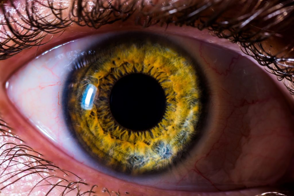
During the eye exam, the doctor will use a slit lamp microscope to view the internal structures of the eye. The microscope can help detect the presence of cells and glitter (increased protein) in the eye fluid, which are hallmarks of uveitis.
Specific laboratory tests and imaging may be necessary to identify the underlying cause of uveitis. These tests are usually guided by the patient's history and clinical findings. For example, a chest X-ray or CT scan may be needed if sarcoidosis is suspected, while an HLA-B27 antigen test may be performed if arthritis is suspected.
Other laboratory tests may include complete blood count, erythrocyte sedimentation rate (ESR), C-reactive protein (CRP), antinuclear antibody (ANA), anti-Sjogren's antibodies, angiotensin-converting enzyme (ACE), and lysozyme levels. The choice of tests is guided by the clinical presentation and the suspected underlying condition.
The differential diagnosis of uveitis is broad, given that it can be a manifestation of several systemic diseases. This includes infectious diseases such as tuberculosis, syphilis or Lyme disease, and autoimmune diseases such as sarcoidosis, Bechchet's disease or ankylosing spondylitis.
However, a significant proportion of uveitis cases are idiopathic, meaning that no cause can be identified despite extensive investigation. In such cases, the treatment is usually aimed at controlling the inflammation and preventing complications.
Additional imaging measures may be required depending on the clinical presentation and suspicion of the underlying disease. For example, angiography with fluorescein (FA) can provide detailed images of the blood vessels of the eye, helping to detect vasculitis, which is often associated with systemic autoimmune diseases such as lupus or Bechette. Optical coherence tomography (OCT) can reveal macular edema, a common complication of uveitis.
In some cases, consultation with other specialists may be necessary. For example, if an infectious cause is suspected, an infectious disease specialist can be consulted. Similarly, if a systemic autoimmune disease is suspected, it may be necessary to involve a rheumatologist in the patient's treatment.
After receiving the diagnosis, appropriate treatment can be started. In autoimmune uveitis, treatment usually involves the use of corticosteroids to reduce inflammation. In cases where an underlying systemic disease is identified, it is also necessary to treat this condition. Immunosuppressive drugs may be used in severe cases or when corticosteroids are ineffective.
In conclusion, the diagnosis and treatment of uveitis require a comprehensive and multidisciplinary approach. Early diagnosis and prompt treatment are essential to reduce the risk of vision loss and improve patient outcomes. Diagnosing uveitis and identifying its underlying cause is a complex process that requires a comprehensive history, physical examination, and focused laboratory and imaging tests. Early diagnosis and quick treatment are essential to prevent irreversible damage and preserve vision.
The author of the article: Dr. Yael Sharon, uveitis specialist
:References
טיפות דמעות הן תמיסות רפואיות או קוסמטיות שמטרתן להקל על
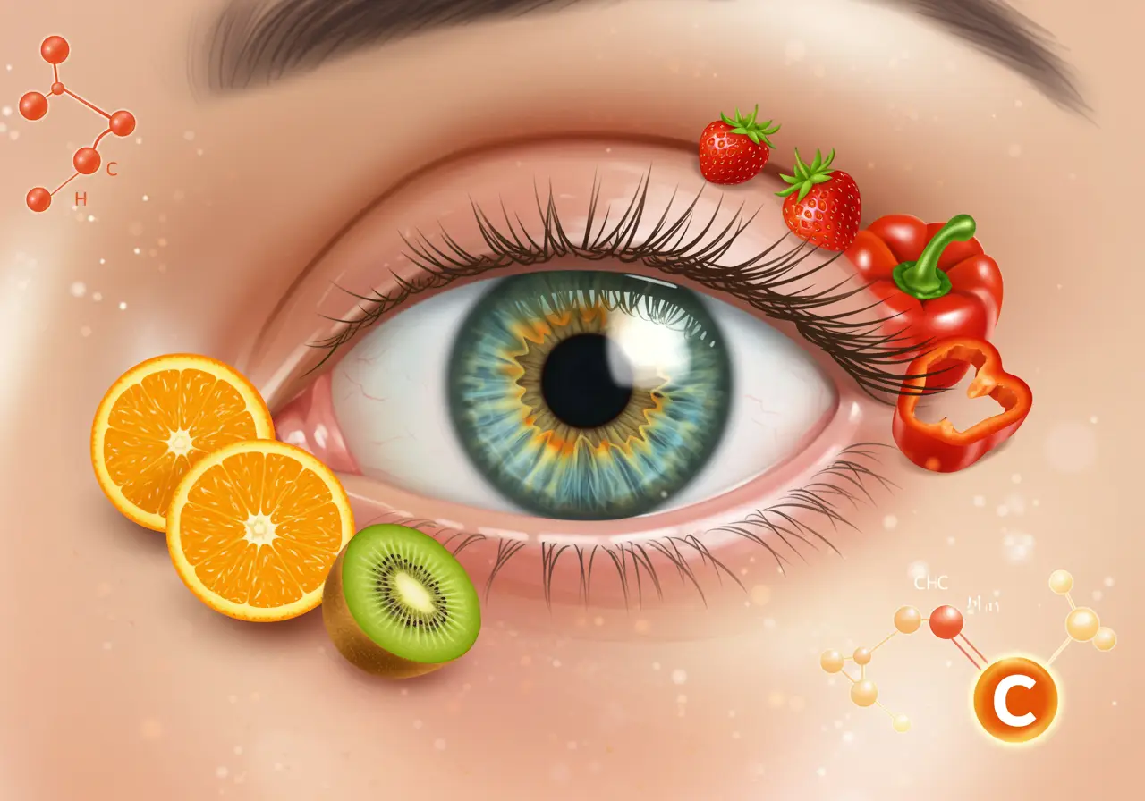
ויטמין C הוא אחד הוויטמינים החיוניים ביותר לבריאות העיניים, הודות